Publish Date: July 2, 2024
Impact of Early Visual Experience On Later Usage of Color Cues
Share this on
How does our recognition ability change when we go from color to grayscale images? The fact that we can perfectly well recognize things in old black and white movies and photographs suggests that there should be little effect of removing color information from an image. Indeed, that is what we find when we test normally sighted children on an object naming task. The children are shown many images, either in color or grayscale, and asked to name the object shown.
The performance of normally sighted children does not change when color information is removed. However, a very different result is seen with newly sighted children born with congenital cataract (Prakash children).
The Prakash children show a decrease in performance when color information is removed from images. In each case, performance with grayscale images is worse than performance with color. We would like to understand why is this? The quest to understand this clinical observation has seeded a basic science investigation. Here is our explanation.
First some background. The first stage of sensing color is the cone photoreceptor in our retina. The brain combines information from three types of cone cells to derive color percepts. Importantly, the cone cells have a developmental progression.
As shown below, a newborn’s cones are very immature and over the course of the first couple of years, they acquire their adult forms. At the beginning of life, the cones are very limited in signaling color information. As the image progression at the bottom shows, a newborn has poor color vision and gains color richness over several months.
A Prakash child, when he or she surgically gains sight at the age of several years, already has mature cones. Thus, right after surgery, a Prakash child begins to sense the visual world in rich color. We might think that this is a good thing. But, counterintuitively, this overly good color perception at the outset may be detrimental for the Prakash child.
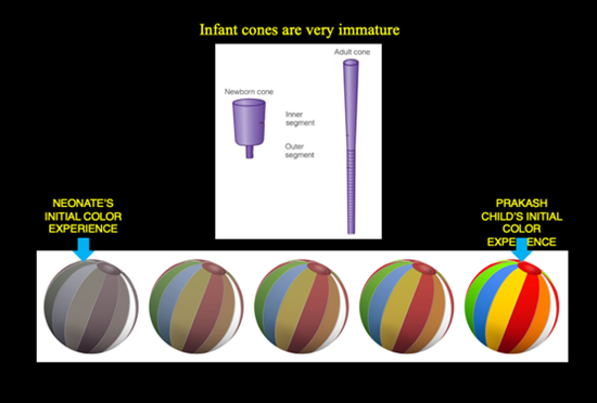
So, to the question of why Prakash children show a significant drop in performance when color information is removed, our hypothesis is that starting with rich color images makes the Prakash child’s visual system overly reliant on color. Hence, the child performs poorly when color is removed (gray-scale images).
Computationally testing the idea that color restriction early in development may lead to greater robustness to color changes later.
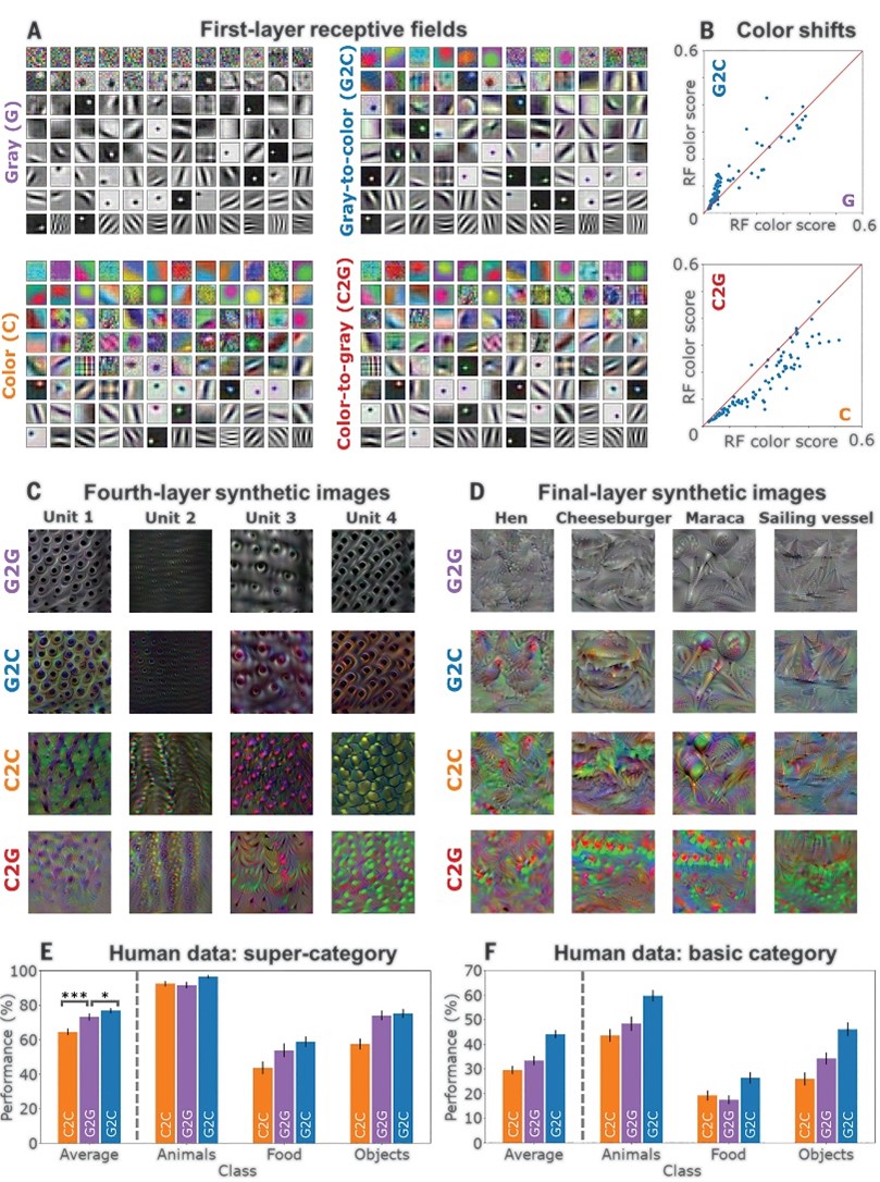
Although this hypothesis nicely explains our clinical data, we want to test it a little more rigorously. We use a computational visual system, specifically a deep network, to examine the impact of different training regimens. In particular, we want to determine if initial color restriction can lead to greater robustness to color changes later.
A network trained only with color images (the C2C regimen) performs poorly when color information is shifted.
However, networks trained on grayscale images, or those that start with grayscale and then see color images have no trouble dealing with color variations. They are robust and their performance is unchanged by color perturbations.
Interestingly, the C2G trained network does not perform as well as the G2C network, demonstrating that the order in which images are shown during training (first grayscale and then color, rather than the other way around) matters for determining final network performance. The biomimetic regimen (gray then color) is better than the non-biomimetic regimen (color then gray). The inference from these results is quite clear.
Both the clinical data from human subjects and the computational data from deep networks suggest that initial experience with achromatic inputs is useful for reducing the reliance on color cues.
“Our findings are significant from three perspectives: 1. They help explain a pattern of deficits observed in newly sighted children, 2. They provide an account for why normal visual development proceeds in the way that it does, and 3. They suggest that the performance of AI vision systems can be improved by incorporating insights from human development”, said Dr. Tapan Gandhi, Professor in Electrical Engineering Department, IIT Delhi and Cadence Chair Professor of Automation & AI.
(The work published in research journal SCIENCE, is a joint collaboration between IIT Delhi; Shroff Charitable Eye Hospital Daryaganj; Dept. of Brain and Cognitive Science, MIT USA; and Project Prakash Charitable Trust, New Delhi. Research paper- https://www.science.org/doi/10.1126/science.adk9587)
Other News
IIT Delhi Researchers Develop High-Speed, Self-Powered Photodetector for Next-Gen Optical Communication
Read More
Atomically thin graphene coating effectively protects glasses from simultaneous mechanical and chemical damage under water: Study
Read More
Sprayable Hydrogel by IIT Delhi Researchers for Improving Wound Healing Exhibits Promising Results in Pre-Clinical Trial
Read More
IIT Delhi Researchers Find a Potential Solution to Develop Perovskite Solar Cells Under Air Ambient Conditions Without Using Anti-Solvents
Read More
IIT Delhi Researchers Develop and Release a Machine Learning and Cloud Computing Based Tool for Rapid and Automated Mapping of Landslide Extent
Read More
IIT Delhi Researchers Demonstrate a New Polymeric Material Having Potential to Develop Advanced Electronic Devices for Data Storage and Encryption
Read More
IIT Delhi Researchers Find a Potential Solution to Regulate Dendrite Growth in Room-Temperature Sodium-Sulfur Batteries
Read More
IIT Delhi Scientists Working on Cure for Brain Cancer Gets Promising Results in Pre-Clinical Trials
Read More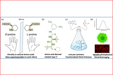
National Centre for Assistive Health Technologies (NCAHT) at IIT Delhi Launches Assistive Technology Products for Visually Challenged
Read More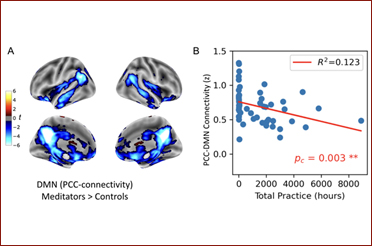
First-Ever Brain Imaging Study on Yoga Nidra Finds Significant Changes in the Brain’s Functional Connectivity during the Practice
Read More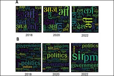
Hinglish Helps Users Engage More Effectively with a Broader Audience On Social Media: Study
Read More
IIT Delhi Researchers Develop Highly Efficient Terahertz Radiation Source for Beyond 6G Technology
Read More

A Study by IIT Delhi Researchers Proposes Solutions for Fair Compensation to Food Delivery Agents in India
Read More
Rural Technology Action Group at IIT Delhi Transfers “AC Motor-Powered Wooden Bead Making Device” Fabrication Technology to Industry
Read More
Researchers Develop Real-Time Bioelectrochemical Sensor for Rapid Water Quality Monitoring
Read More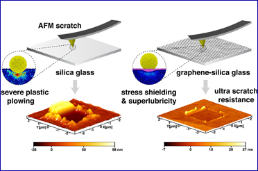
IIT Delhi Researchers Find Ultrathin Graphene Coating Can Make Glasses Extremely Scratch-Resistant
Read More
Researchers and Practitioners Propose Creation of a New Mission Energy Access Programme to Support Global Goal of Ending Energy Poverty by 2030
Read More
Researchers to Global South: Use Emerging Technologies and Strengthen International Collaboration to Overcome Challenges in Adapting to Climate Change
Read More
IIT Delhi Researchers Develop Scalable Wearable Pressure Sensor That Can Help Doctors and Specialists Analyze Gait Patterns and Postural Deformities
Read More
Researchers at IIT Delhi Achieve Trusted-node-free Secure Quantum Communication for 380 km in Standard Telecom Fiber
Read More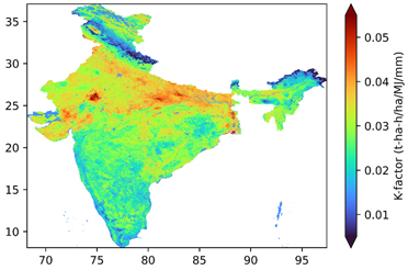

IIT Delhi's Exoskeleton Device Heads for International Footprint to Australia for Clinical Trials in Collaboration with Proxmed
Read More
Researchers Develop a Simulation Tool for Planning Operational Response During a Health Crisis
Read More
AI/ML Model Developed by IIT Delhi-led Researcher Predicts 2023 to be a Normal Monsoon Year
Read More
Researchers at IIT Delhi Develop Mobile Robot “Robomuse 5.0” Capable of Carrying Payloads Up to 100 kg
Read More
Evidence of Structural Brain Plasticity Observed Following Treatment to Congenital Blind Humans
Read More
Researchers at IIT Delhi and IIT Bombay Develop Highly Efficient Spintronics-Based Neuromorphic Hardware
Read More
Researchers' Efforts to Develop a Next-Generation Vaccine Against COVID-19 Give Promising Results in Animal Trial Phase
Read More
Researchers Led by IIT Delhi Scientist Develop a VLP-based Vaccine Candidate Against COVID-19
Read More
IIT Delhi Showcases Technologies with Societal Impact, Innovations in Clean Energy, Healthcare, Manufactuting at All IITs R&D Fair IInvenTiv
Read More
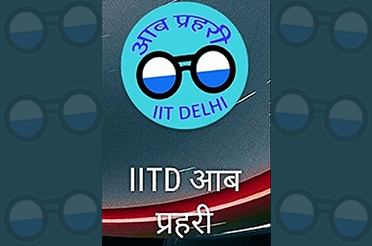
IIT Delhi Launches Mobile Application ‘IITD Aab Prahari (आब प्रहरी)’ to Address Waterlogging Issues in Urban Areas During Monsoon
Read More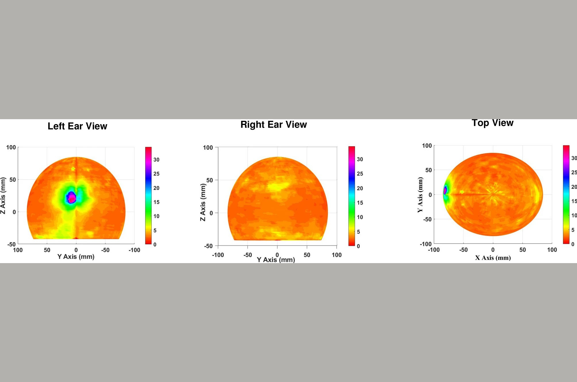
IIT Delhi Researchers Propose Non-invasive, Time Efficient and Patient Friendly Diagnostic Tool for Epileptogenic Zone Detection
Read More
Researchers at IIT Delhi Develop Map to Highlight Areas Prone to Rainfall-induced Erosion in India
Read More
Researchers at IIT Delhi Demonstrate How Polyarylquinone Molecule Can be Synthesized Easily
Read More
IIT Delhi Researchers Develop Low-Cost Buckling Restrained Braces That Can Improve Earthquake Resistance of Structures
Read More
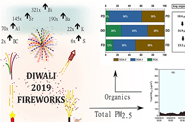
Biomass Burning Drives Poor Air Quality in Delhi Post Diwali, Not Fireworks: IIT Delhi Study
Read More
Researchers Led by IIT Delhi Develop Technology to Enable Use of Environment-Friendly Dimethyl Ether as Fuel in Automotive Vehicles
Read More
IIT Delhi Researchers Design and Demonstrate a New Strategy for Development of Drug Molecules
Read More
IIT Delhi Researchers Develop High Efficiency, Shadow-less, Portable Solar PV Towers for Power Generation
Read More

RT-PCR-based Assay for Identification of Omicron Variant of SARS-CoV-2 Developed at IIT Delhi
Read More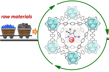
IIT Delhi Researchers Develop Catalytic Technology for Sustainable Production of Chiral Active Pharmaceutical Ingredients
Read More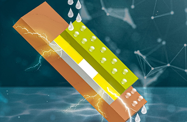

IIT Delhi, RDSO Researchers Develop Easy to Use Train Simulation Software ‘Runtrain#’ to Help in Train Timetabling Methods
Read More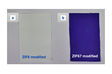
IIT Delhi Researchers Develop Modified Cotton Fabric Capable of Adsorbing Harmful Air Pollutants from Air
Read More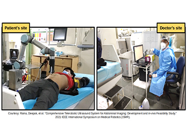
IIT Delhi, AIIMS New Delhi and Addverb Co-develop Telerobotic Ultrasound System During COVID Times
Read More
IIT Delhi Researcher in Collaboration with NUS Designs Device for High Density Magnetic Memory
Read More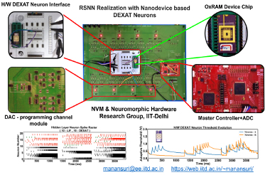
IIT Delhi Researchers Demonstrate a New Brain-inspired Artificial Neuron for Building Accurate and Efficient Neuromorphic AI Systems
Read More
IIT Delhi Scientist led Research Team Develops Novel Antifungal Strategy for Fungal Eye Infection
Read More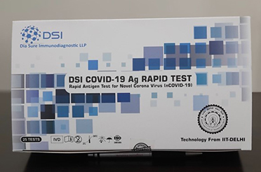
Minister of State for Education Shri Sanjay Dhotre launches the Rapid Antigen Test Kit for COVID-19 developed by IIT Delhi
Read More
Technology for Hydrogen Utilization in Spark-Ignition Engine Generator for Electricity Generation with Zero-Emission Developed by IIT Delhi Researchers
Read More